“I hear of none that makes any other Use of that Instrument but for Diversion and Pastime.”
Robert Hooke, 1665
While there were many botanists and zoologists who used microscopes before the 19th century, there were few physicians. The red blood cell was described in 1667 by Jan Swammerdam (1637–1680) and in 1673 by Marcello Malpighi (1628–1694). Malpighi also proved Harvey’s observation that blood circulated around the body by identifying capillaries connecting the arteries and veins. The microscopic observations of Anton Leeuwenhoek (1632–1723), a Dutch drapery maker, excelled because of his skill in making high quality lenses. He first illustrated red blood cells in his Arcana in 1695. He wrote many letters to the Royal Society in London giving descriptions and illustrations of bacteria from the human mouth, protozoa, spermatozoa, striations of skeletal muscles and epithelial cells from a wart on the trunk of an elephant in the Amsterdam Zoo. Despite these early observations, microscopes were slow to be adopted for clinical use.
Why did 18th century doctors not use microscopes in their work?
They generally believed:
Disease was caused by an imbalance of humours and external miasmas.
Microscopic studies diverted physicians from making bedside observations.
Nature’s inner structures were too complex to understand.
There was no logical relationship between the macroscopic and microscopic world.
Although instruments using multiple lenses to increase magnification were soon developed, these compound microscopes were not capable of producing good images until 1830 when Joseph Jackson Lister, father of the surgeon Joseph Lister, introduced lenses that eliminated blurring and colour distortion, which had previously been a problem with higher power microscopes.
Lister’s breakthrough, the ‘achromatic’ lens, transformed the microscope into a powerful tool capable of much higher magnification. Binocular eyepieces made viewing more comfortable and differential staining techniques facilitated better identification of microbes and human tissues.
These improvements in microscopic technology converged with the emergence of bacteriology driven by the work of Louis Pasteur and Robert Koch. By the 1880s, the microscope became an essential tool of doctors in the daily practice of identifying pathogens. This pioneering work allowed easy identification of epidemic and endemic diseases. Doctors could now finally see what caused infectious disease and could combat its spread through quarantine, disinfection, vaccines, and antibiotics. However, there was one class of micro-organism that still remained invisible to them, even with the most powerful optical instruments.
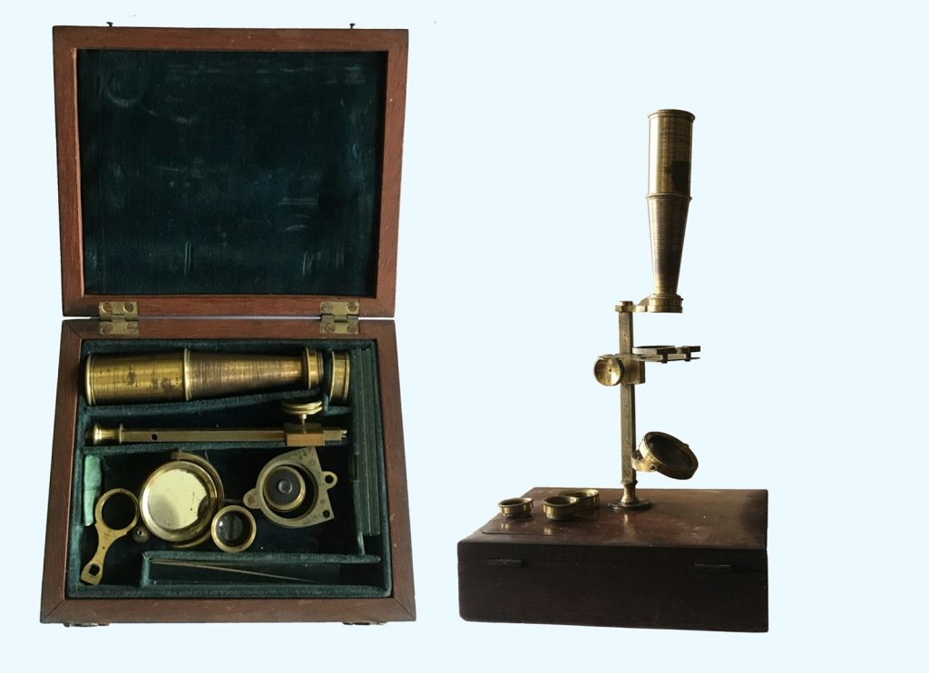
Portable microscope designed by Charles Gould, made c1830 by G & C Dixey, London). Similar microscopes were made by the Bath optician and instrument maker Jacob Abraham (1772-1845)
The microscope could be dissembled and transported in its mahogany case. It was rarely used for medical work. (BMM collection).
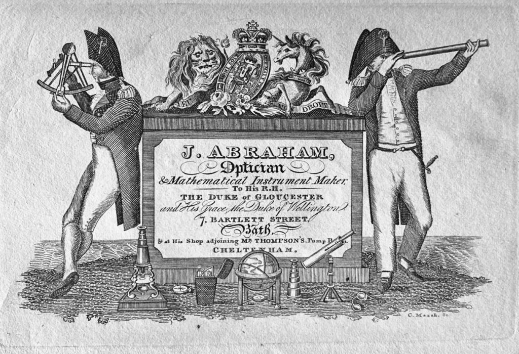
Advertising engraving for Jacob Abraham. (Private Collection, on loan to BMM).
Fifty million times magnification
Although improvements in the design of lenses enabled doctors to see smaller objects in greater detail, very tiny objects like viruses remained invisible because their dimensions were smaller than the wavelength of light. This was overcome by using an electron beam, which has a much shorter wavelength than visible light. Electron microscopes allowed viruses and other minute structures to be seen in great detail. The first electron microscopes were developed in the 1930s and the first commercial electron microscopes appeared in 1938 made by the German electrical engineering company, Siemens. This type of microscope is known as a transmission electron microscope or TEM. Modern TEMs can form images of structures as small as 0.5 Å which represents a magnification of 50 million times. The main disadvantage of a TEM is a requirement for specimens to be prepared in extremely thin sections. Creating such thin specimens for biological and medical work is very challenging. This has been solved by the development of the scanning electron microscope or SEM and, more recently, an electron microscope that combines scanning and transmission technique (STEM). The high-resolution three-dimensional images which these instruments can produce have opened up an entirely new world of sub-microscopic structures.
The Bath Microscopical Society was set up in 1858 and continued until around the end of the century when the membership had dwindled to a single figure. Monthly meetings were held in the Royal Mineral Water Hospital. Although the society had a number of medical members, including Dr Randle Wilbraham Falconer who was its president in 1864, interest lay in microscopical examination of biological and geological specimens rather than clinical material. There was also a short-lived Ladies Microscopical Society founded by Miss Emily Jarrett of Camerton Court in 1897. It functioned until 1911 when Miss Jarrett died.